What is Laser Lithotripsy? | Laser Stone Surgery in South Bopal | Laser Stone Removal in South Bopal | Stone Disease Treatment in South Bopal Ahmedabad
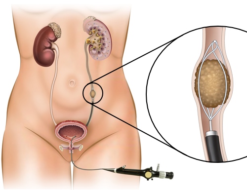
Urinary stones are a common source of discomfort and in many cases require a surgery for cure. Till 1980’s this surgery was done by open method which was morbid. After 1980’s endoscopic surgery became the standard of care. The endoscope is introduced in the urinary tract and once the stone is accessed it has to be fragmented and the fragments are then removed from the urinary tract.
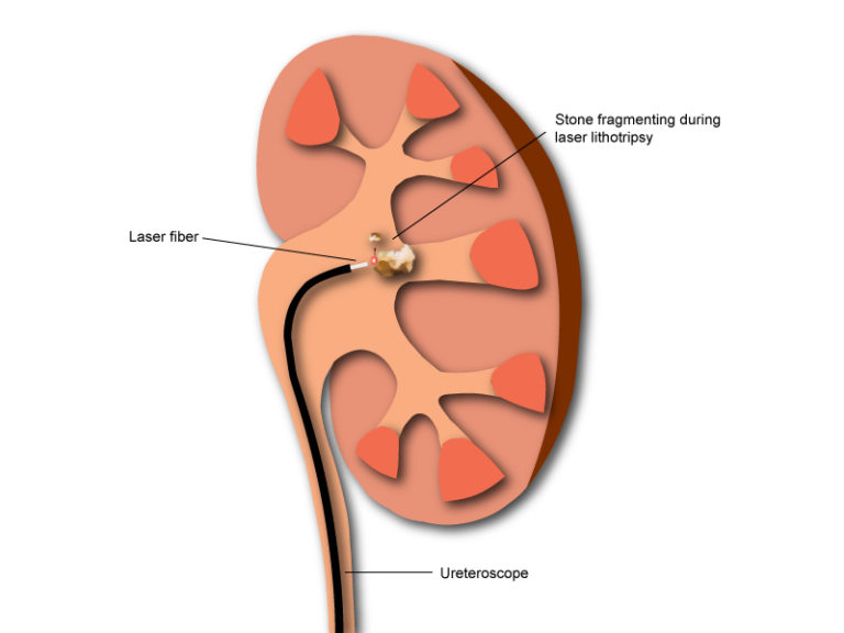
Many people think that endoscopic surgery and laser surgery are the same. But this is not the case. Endoscopic surgery is a minimally invasive method of treating urinary stone. Once the endoscope reaches the stone it can be fragmented by various methods like laser, lithoclast, ultrasonic machine etc. But out of these laser is the most effective and also the safest method. Laser lithotripsy has many advantages over other methods of stone fragmentation as below

1. Renal Calculi
Till 1990’ open surgery was the procedure of choice for treatment of renal calculi. But this carried high morbidity due to the large incision, pain, late recovery and bad cosmetic result.
Today renal stones are treatment by minimally invasive methods. The three modalities of treatment of renal stones are ESWL, RIRS and PCNL. Open surgery for kidney stone is almost never required.
A. Extracorporeal Shock Wave Lithotripsy (ESWL)

B. Retrograde Intrarenal Surgery (RIRS)

Renal stones < 2 cm sometimes may be unsuitable for ESWL. These can be treated by Retrograde Intrarenal Surgery (RIRS) in which a flexible ureteroscope is inserted through the urethra into the ureter. This can then access the calyces of the kidneys since it has a flexible tip. The stone once located can then be fragmented with laser. RIRS needs exclusive use of laser since the laser fibers are also flexible and can bend along with the scope.
Patients with abnormal renal anatomy (eg. horseshoe kidney, ectopic kidney) sometimes develop renal stones. RIRS is an excellent option for these patients since it can bypass the difficulties offered by the abnormal anatomy.
C. Percutaneous Nephrolithotomy (PCNL)
Advantages of PCNL:
2. Ureteric Calculi
What is Flexible Ureteroscopy/Retrograde Intrarenal Surgery (RIRS)?
Ureter is a tube that carries urine form kidney to the urinary bladder. Many urinary stones get trapped in the ureter and cause obstruction to the urine flow. This can lead to excruciating pain. Many such patients need to undergo surgery. In today’s era this surgery is done with ureteroscope. Ureteroscope are of two types rigid and flexible. Rigid urteroscope many times cannot access the upper ureter and also cannot access the calyces of the kidney. A flexible ureteroscope does not have these limitations and can access the upper urinary tract with ease.
Lihtotripy with flexible ureteroscope is called Retrograde Intrarenal Surgery (RIRS). RIRS needs exclusive use of laser since the laser fibers are also flexible and can bend along with the scope. Many upper ureteric stones migrate into the kidney during fragmentation and cannot be accessed by a rigid ureteroscope. These migrated stones then need additional treatment like ESWL or PCNL for clearance. Flexible ureteroscope can clear these stones transurethrally in the same sitting without an incision.
Many patients have abnormal anatomy of the kidney or ureter which limits the use of rigid scopes. Flexible ureteroscope can bypass these difficulties of abnormal anatomy of the upper urinary tract eg. horseshoe kidney, ectopic kidney.

Advantages of URSL/RIRS :
Important factors to consider while deciding treatment for ureteric stones.
1. Stone Burden (ie. size & no) Width of the stone is the most imp factor affecting the likelihood of stone passage. 5-mm is the breakpoint. For stones < 5 mm, conservative management is considered. For ureteric stones greater than 5mm URSL is recommended
2. Duration of Stone Presence any ureteric stone has the potential to cause irreversible loss of renal function. Symptoms and stone size don’t predict loss of renal function. For stones > 5mm patient should be properly counseled about the risks associated with waiting. If the stone is small it is safe to wait for 2-3 weeks provided the degree of backpressure changes are minimal.
3. Pain Majority of the patients present with pain in the flank which occurs due to obstruction to the upper urinary tract. Pain not getting controlled with oral analgesics is an indication for URSL.
4. Infection associated with ureteric stones is potentially life threatening and a urologic emergency. Patients may have fever, renal angle tenderness or may have signs of septic shock eg hypotension. Urgent drainage of obstructed upper urinary tract by ureteric stenting or by a nephrostomy tube is required in such cases. Appropraite antibiotics should be started. Definitive treatment of the stone is delayed till urine cultures are negative and patients has recovered completely from the infection.
5. Solitary Kidney: Ureteral stones in surgically/functionally solitary kidney demands prompt treatment to prevent renal failure.
3. Urinary Bladder Calculi
Etiology
Disease processes resulting in urinary stasis in the bladder eg. due to enlarged prostate, stricture urethra, bladder diverticulum or neurogenic problem eg spinal cord injury. They may also be due to the introduction of a foreign body in the bladder eg. a non absorbable suture used in a previous surgery. Characteristic symptoms are Dysuria, Intermittency & Hematuria. Other symptoms which may be present are Freq, reduced stream, lower abdominal pain. Diagnosis requires an X ray KUB & USG on full bladder.
Treatment
The primary goal of surgical stone management is to achieve maximum stone clearance with minimal morbidity. Treatment of bladder calculi depends on the size of the stone with 3 cm being the cut off. For stones < 3 cm Transurethral cystolitholapaxy is recommended. The picture above shows a Mauer Mayer stone punch which is used to crush the stone. This procedure is not recommended for larger stones as the procedure then takes a long time, increasing the future risk of a urethral stricture.
For large bladder stone ie > 3cm options are either an percutaneous/Open cystolithotomy.
Per Cutaneous Cystolithotomy (PCCL) is a minimally invasive technique which involves creation & dilatation of a suprapubic tract in a distended bladder. An Amplatz sheath is used to maintain the tract. A nephroscope is passed through the sheath & stone is fragmented using pneumatic lithoclast. A suprapubic catheter is kept after the surgery & pt is discharged the next day with the catheter which is removed after 3-4 days. This technique eliminates the risk of trauma to the urethra from repeated passages of instruments in case of a large stone.

Open cystolithotomy is another option for a large vesical stone but it is not favoured due to the need of prolonged catheterization, increased length of hosp stay, longer time for recovery and poor cosmesis. It is done only if the stone is very large and not amenable for Per cutaneous cystolithotomy (PCCL)

The most important thing is to treat the cause of bladder stone formation to avoid recurrence. This may be a
Risk of Recurrence
First time stone formers have a 50% risk of recurrence within the next 10 yrs. Hence all of them are provided empiric fluid & dietary recommendations as below
